WEST LAFAYETTE, Ind. – Purdue University researchers are developing a novel biomedical imaging system that combines optical and ultrasound technology to improve diagnosis of life-threatening diseases.
Photoacoustic tomography is a noninvasive technique that works by converting absorbed optical energy into acoustic signal. Pulsed light is sent into body tissue, creating a small increase in temperature that causes the tissue to expand and create an acoustic response that can be detected by an ultrasound transducer. The ultrasound data is used to visualize the tissue.
“The nice thing about photoacoustic tomography is the compositional information,” said Craig Goergen, an assistant professor in Purdue’s Weldon School of Biomedical Engineering. “It provides information about where blood and lipid are located, along with other essential information.”
The ultimate goal is to enhance the clinical care of patients.
The results of a study describing an adjustable photoacoustic probe with improved light delivery and image quality were published Tuesday (Aug. 28) in the journal Photoacoustics.
The system provides real-time compositional information of body tissue without the need for contrast agents and with better depth penetration compared with conventional optical techniques.
Photoacoustic tomography can be used to detect or monitor a myriad of diseases, including cardiovascular disease, diabetes, and cancer. Those are diseases that the Centers for Disease Control and Prevention lists as among the most common, costly, and preventable of all health problems. Heart disease and cancer each account for one in every four deaths a year in the United States, and more than 30 million Americans, or more than 9 percent of the population, have diabetes. The cost of those three diseases a year in the United States is more than $718 billion a year, according to the CDC.
“That means there will be a great need for medical imaging. Trying to diagnose these diseases at an earlier time can lead to improved patient care,” Goergen said. “We are in the process now of trying to use this enhanced imaging approach to a variety of different applications to see what it can be used for.”

Among other potential uses for photoacoustic tomography is the mapping of lipid deposition within an arterial wall that can cause other health problems, measuring cardiac tissue damage and tumor biopsies. Using photoacoustic tomography for intraoperative tumor biopsies could help surgeons make sure they remove all the cancer from a patient, Goergen said.
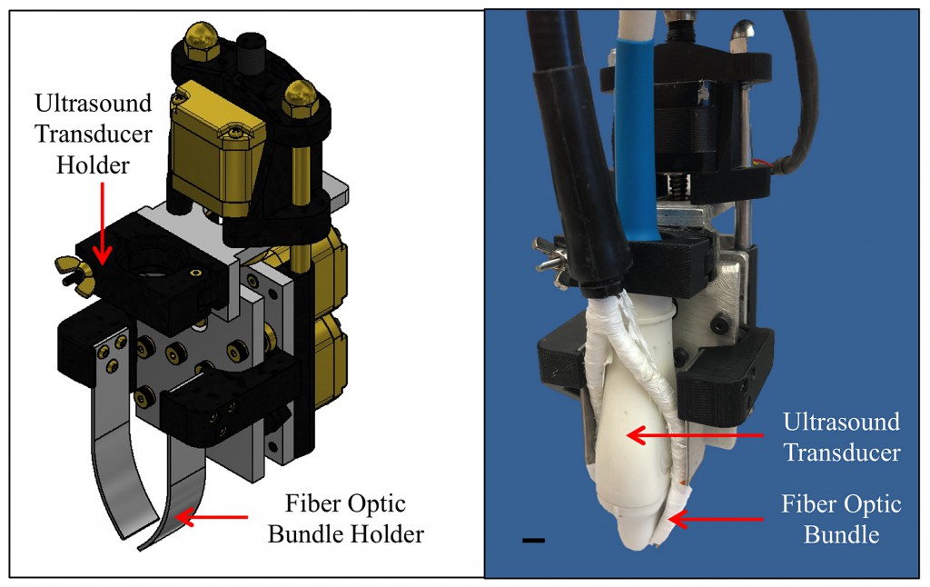
One of the challenges of photoacoustic tomography is improving the penetration depth and signal-to-noise ratio past optical absorbers. The researchers believe creating optical manipulation techniques to maximize photon density could provide a solution. As a result, they have created a motorized photoacoustic holder that allows users to easily maneuver the aim of the device and tune the depth where light is focused, improving the light penetration depth and signal-to-noise ratio.
A video about the acoustic tomography is available at https://bit.ly/2yJddb0. A complete list of co-authors is available in the abstract. The research has been funded by the National Institutes of Health.
The Purdue researchers are interested in talking with other companies about other possible uses for photoacoustic tomography. The researchers have a patent pending for the technology with the help of the Purdue Office of Technology Commercialization.
About Purdue Office of Technology Commercialization
The Purdue Office of Technology Commercialization operates one of the most comprehensive technology transfer programs among leading research universities in the U.S. Services provided by this office support the economic development initiatives of Purdue University and benefit the university’s academic activities. The office is managed by the Purdue Research Foundation, which received the 2016 Innovation and Economic Prosperity Universities Award for Innovation from the Association of Public and Land-grant Universities. For more information about funding and investment opportunities in startups based on a Purdue innovation, contact the Purdue Foundry at foundry@prf.org. For more information on licensing a Purdue innovation, contact the Office of Technology Commercialization at innovation@prf.org
The article is available at: https://doi.org/10.1016/j.pacs.2018.08.002
Abstract
One cause for suboptimal photoacoustic tomography (PAT) penetration depth is attenuation of incident light by soft tissue. To better understand this problem, we investigated the effects of illumination fiber optic bundle geometry on PAT penetration depth and signal-to-noise ratio. An adjustable, motorized PAT probe was used to reduce probe-skin reflection artifacts and improve light distribution in the image acquisition plane by tuning fiber orientation. We validated our motorized PAT probe through Monte Carlo simulations and ex vivo imaging of a tissue mimicking phantom, and in vivo imaging of murine periaortic fat. Overall, our ex vivo results showed a several millimeter improvement in penetration depth and in vivo results showed a >62% increase in lipid signal-to-noise ratio. Our PAT probe also utilized a 7-μm aluminum filter to block in vivo probe-skin reflection artifacts. Together, these findings showed the importance of optimizing illumination geometry to enhance PAT image quality.
Source
Purdue, press release, 2018-08-28.
Supplier
Share
Renewable Carbon News – Daily Newsletter
Subscribe to our daily email newsletter – the world's leading newsletter on renewable materials and chemicals